Introduction
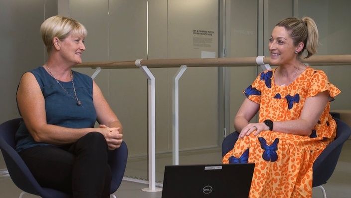
The Athletic Ankle Masterclass, taught by Dr Ebonie Rio and Dr Sue Mayes, provides a thorough overview of how to accurately assess and manage various ankle pathologies including Posterior Ankle Impingement and tendon pain. This Executive Summary provides clinically relevant, actionable information that may be useful to health professionals.
Part 1: Tendon Pain
Tendon pain, typically of the achilles for ankle pathologies, can be difficult to diagnose and manage. Differentiating between tendinopathy, peri-tendon, bursae, and tendon ruptures is crucial as each condition is aggravated by different loading types, and subsequently require a slightly different treatment approach to appropriately manage it. Scans are often utilised to help with diagnosis, however their utility for tendon pain is questionable given the high presence of incidental findings.
- The language that clinicians choose to use to describe pathologies is crucial (e.g., tendonitis vs tendinopathy) as it can significantly affect perspectives and expectations for rehabilitation
- Key characteristics of a tendinopathy include a) Localised pain to the tendon, b) aggravated by dose (rate) dependent load, c) symptoms improve once warmed up and d) symptoms generally reveal themselves 24 hours after an inciting event.
- Tendinopathy is not an inflammatory condition.
Part 2: Posterior Ankle Pain
Our understanding of Posterior Ankle Impingement Syndrome (PAIS) continues to evolve. The condition was previously attributed to the presence of an Os trigonum, a failed fusion of part of the talus, however we now know there is a high prevalence of this finding in asymptomatic individuals. Several other factors are now thought to contribute to this presentation, such as flexor hallucis longus (FHL) tenosynovitis or subtalar joint instability.
- PAIS should be thought of as a syndrome, as it can involve impingement or irritation of cartilage, bone, Kager's fat pad, FHL, synovium, the joint capsule, and ligaments.
- Subtalar joint instability, often due to a previous ankle sprain, can lead to subtalar joint or talocrural synovitis and contribute to PAIS.
- FHL Tenosynovitis often occurs due to repetitive movement through full range of motion (ROM) of the big toe and talocrural joint. Weak calf muscles contribute to its development.
Masterclass Preview
Plyometrics are essesntial to a successful ankle and Achilles tendon rehab plan.
Enjoy this free preview showing how Ebonie and Sue incorporate plyometric exercises into their rehab plans with their athletes.
Part 3: Clinical Assessment of the Foot and Ankle
There is a plethora of structures crammed into the ankle and proximal foot that may play a part in the development of ankle pathologies. As such, a detailed and thorough clinical assessment is paramount to ensure an accurate diagnosis.
- Pain location is an incredibly useful tool to differentiate between the peritenon, insertional or mid portion achilles tendinopathy, sural nerve, plantaris and PAIS.
- A graded loading assessment, progressing from a double leg calf raise to eventually a single leg hop for maximal height, helps to isolate the source and severity of the pain, therefore aiding with diagnosis.
- Special tests should be performed to assess the ROM of the big toe and subtalar joint, to check for signs of PAIS, and to palpate the sinus tarsi and FHL which may be contributing to an individual’s presentation.
Part 4: Rehabilitation of the Foot and Ankle
Exercise is a valuable tool in foot and ankle rehabilitation. However, it can be difficult to apply rehabilitation principles when faced with a range of pathologies which each have their own quirks and intricacies. Understanding the type of load that aggravates the individual’s condition is crucial, as we can then remove or temporarily reduce that provocation in rehabilitation. Beyond this, knowing which muscles and exercises to target is a further challenge. Optimal rehabilitation of the foot and ankle should focus on the calf, intrinsic muscles of the foot and the rest of the kinetic chain.
- Single leg calf raises are a fantastic exercise, and individuals should aim for at least 25 repetitions with correct alignment of the foot, full height, knee straight and limit use of momentum to maximally load the calf complex.
- The body works as individual limbs and so our rehabilitation must reflect this. Double leg activities lead to compensation from other muscles and joints.
- Stair climbing is a great way to introduce plyometrics for the calf and intrinsic muscles of the foot, and is relevant for a wide range of the general population.


